Five decades after Lennart Nilsson’s historic photo essay on the human foetus came out, the argument around fetuses, embryos, abortion, reproductive rights, etc. has oscillated from religion to politics. But back in 1965, when times were simpler, they mainly talked about how he could achieve this feat.
These incredible photographs were taken with conventional cameras with macro lenses, an endoscope and scanning electron microscope. Nilsson used a magnification of hundreds of thousands and “worked” right in the womb.
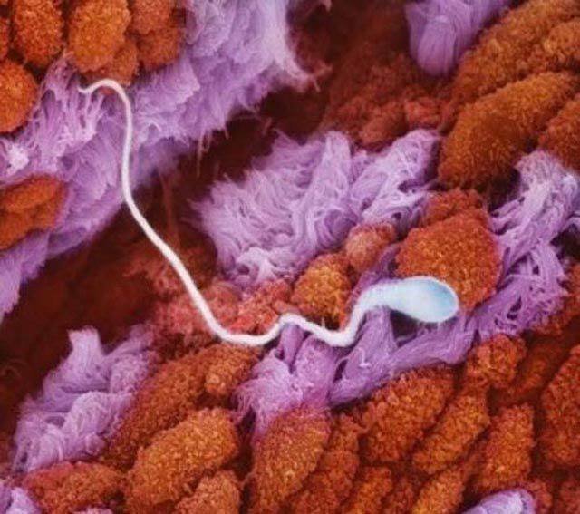
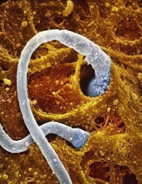
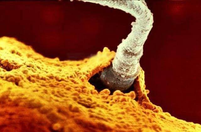
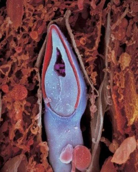
Sperm in the Fallopian Tube
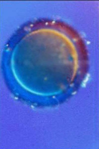
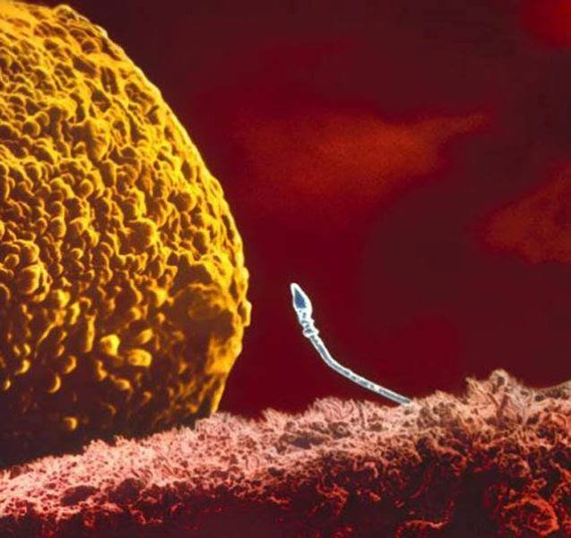
An egg cell
ADVERTISEMENT
ADVERTISEMENT
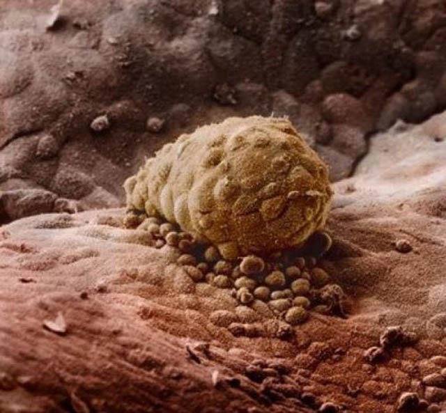
The sperm 5-6 days.The clump has developed into a blastocyst, containing many more cells,and has entered the womb.

8 days. The human embryo is attached to a wall of the uterus.
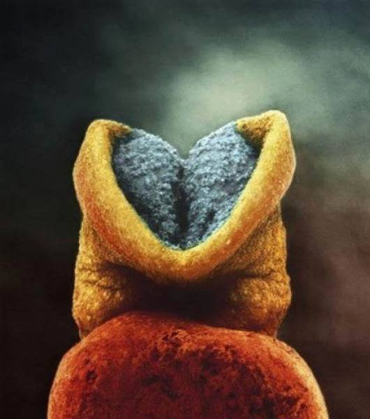
The brain starts to develop in the human embryo.
ADVERTISEMENT
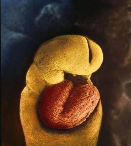
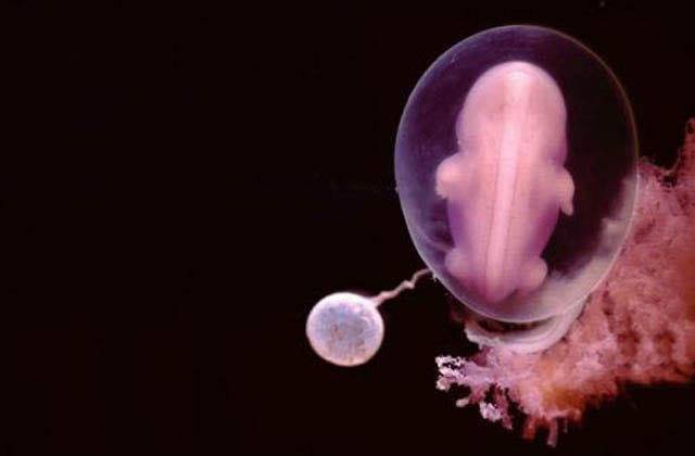
24 days.The one-month-old embryo has no skeleton yet.There is only a heart that starts beating on the 18th day.
At 4 Weeks
5 weeks. Approximately 9 mm.
ADVERTISEMENT

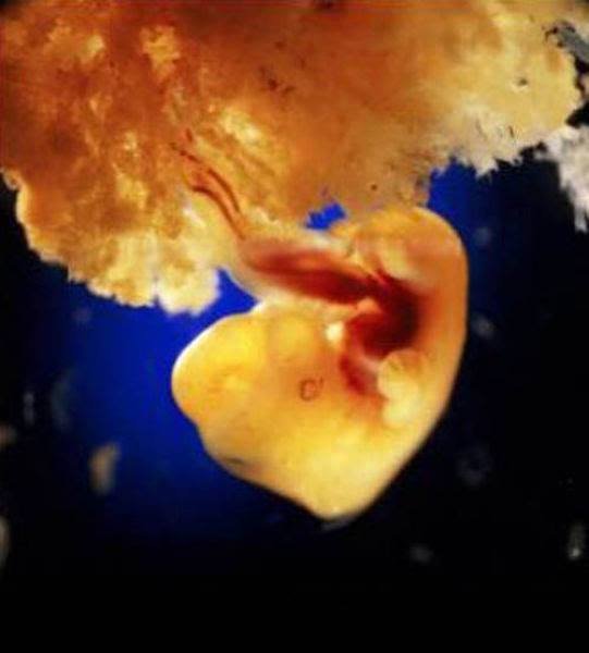
40 days. Embryonic cells form the placenta. This organ connects the embryo to the uterine wall allowing nutrient uptake,waste elimination and gas exchange via the woman’s blood supply.
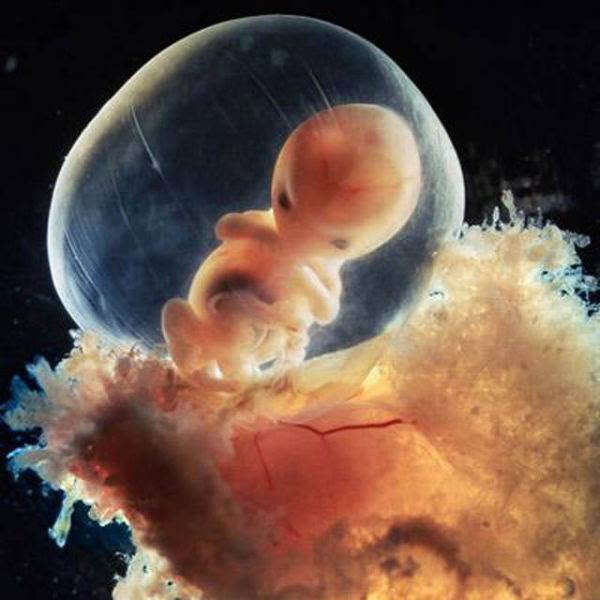
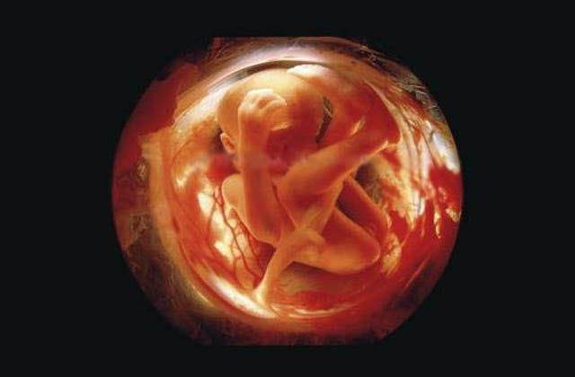
8 weeks. The rapidly-growing embryo is well protected in the foetal sac.
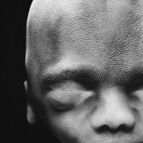
10 weeks. The eyelids are semi-shut. They will close completely in a few days.
ADVERTISEMENT
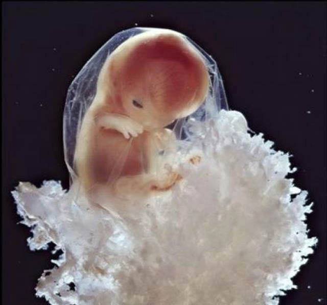
16 weeks. The foetus uses its hands to explore its own body and its surroundings.
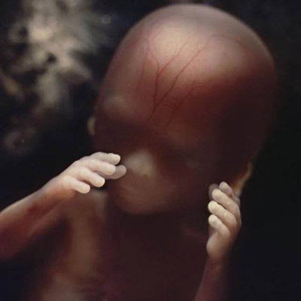
The skeleton consists mainly of flexible cartridge.A network of blood vessels is visible through the thin skin.
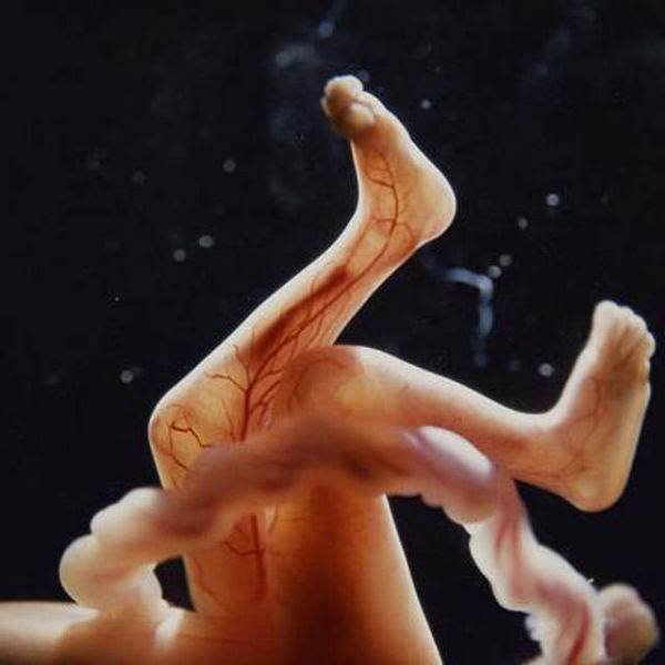
18 weeks. Approximately 14 cm. The foetus can now perceive sounds from the outside world.
ADVERTISEMENT

19 Weeks

20 weeks. Approximately 20 cm. Woolly hair, known as lanugo, covers the entire head.
24 weeks
ADVERTISEMENT

26 Weeks
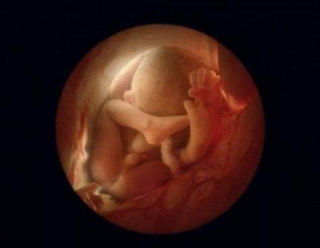
6 months. The little human is getting ready to leave the uterus. It turns upside down because it will be easier to get out this way.

36 weeks. The child will see the world in 4 weeks.
ADVERTISEMENT
You can check out all of Lennart Nilsson’s work here .
h/t India TV
Top picks for you