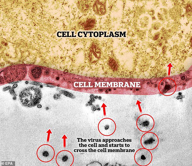
Latest images of the exact moment when the coronavirus infects a cell have been captured by scientists at Latin America’s largest medical research center by using a powerful electron microscope.
These pictures have been taken by experts at the Oswaldo Cruz Foundation (Fiocruz) in Brazil while they studied the replication of the coronavirus.
Produzidas durante estudo no Instituto Oswaldo Cruz @fiocruz, imagens inéditas no Brasil mostram o exato momento da infecção do novo coronavírus em célula #Covid19 https://t.co/PzALusDwep pic.twitter.com/sbVHVcgq3k
— Agência Fiocruz (@agencia_fiocruz) April 8, 2020
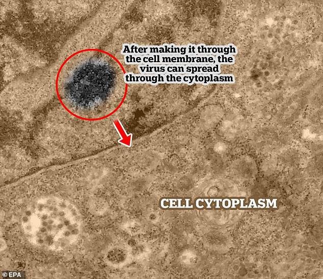
These images which were taken by a transmission electron microscope showed how a series of dark points that are actually viral particles of the pathogen which can magnify objects up to two million times their normal size.
This is the first part of the infection process. The block dots (the coronavirus) can be seen making their way into the cell membrane
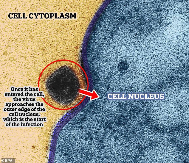
The virus now is entering the cell nucleus – where the genetic material for the cell is kept.
Here, this black dots have infected the cell’s cytoplasm.
The experts at the centre said in a statement that the cells used were not human. These cells were originally isolated from the African green monkey and are regularly used in cell cultures in laboratories.